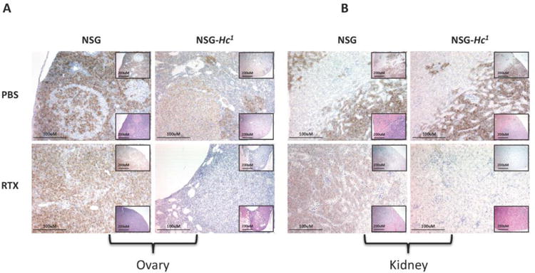
Figure 4.

Representative images of the (A) ovary and (B) kidney 38 days post-engraftment displaying level of Daudi cell engraftment (brown stained cells) in different cohorts of age- and sex-matched rituximab-treated NSG and NSG-Hc1 mice. Low-magnification images at top right showing anti-CD45 staining IHC, at bottom right showing H&E staining, (×10, scale bar = 200um) showing; high-magnification image (×20, scale bar = 100um) showing anti-human CD45 IHC. PBS, phosphate buffer saline; RTX, rituximab. There were also increased numbers of Daudi cells in the livers, spleens, lungs, and lymph nodes of NSG mice (data not shown).
