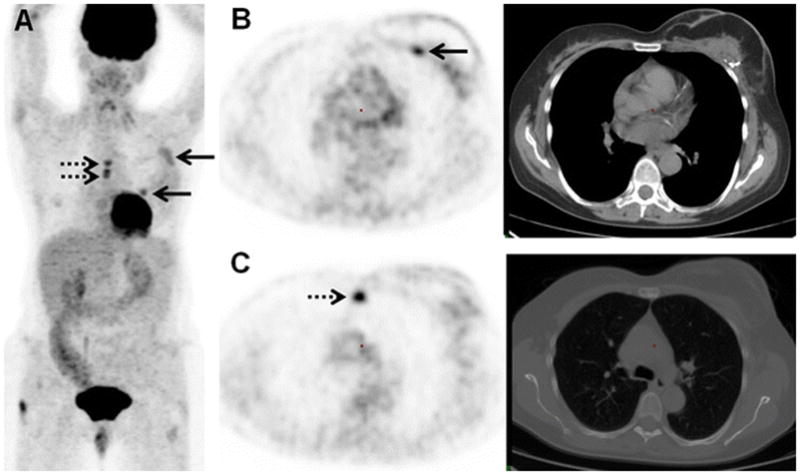
FIGURE 3.

61-year-old woman with initial stage IIB TNBC upstaged to stage IV on 18F-FDG-PET/CT. (A) 18F-FDG MIP image demonstrates the 18F-FDG-avidity overlying the breast breast surgical bed (arrows) and midline chest (dashes arrows). (B) Axial PET and axial CT image on soft tissue window demonstrate the FDG-avid post-surgical changes association with mastectomy and reconstruction 2 weeks before 18F-FDG-PET/CT (arrow). (C) Axial PET and CT image on bone window localize 18F-FDG-avid foci to the sternum without clear CT correlate (dashes arrow), subsequently biopsy-proven to be an osseous metastasis.
