Figure 2.

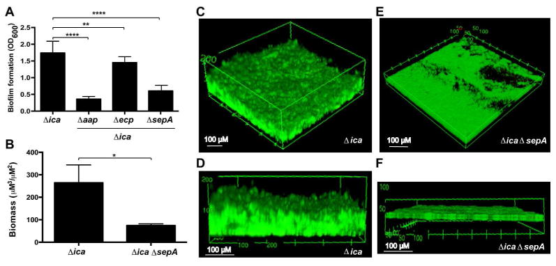
Mutation of the sepA metalloprotease results in decreased PIA-independent biofilm formation. A. Microtiter plate biofilm formation. Strains were grown statically for 20 hours in microtiter plates and the biofilm biomass was stained with crystal violet. Biofilm formation is shown as the absorbance at 600 nm of the solubilized crystal violet. Results are pooled from three experiments with three replicates each. One-way ANOVA with multiple comparisons (Bonferroni correction) was performed. ** indicates p < 0.01; **** p < 0.0001. B. COMSTAT2 results quantifying total biomass in flow cell images. Biomass in live and dead channels was quantified from three images per strain and combined. Total biomass is reported as volume per square μM. * indicates p < 0.05. C-F. Flow cell biofilm formation. Strains were grown under flowed media for 40 hours and stained with the BacLight live:dead kit. Angled (C) and side (D) views of representative images from 1457Δica at 20× magnification are shown. Angled (E) and side (F) views of representative images from 1457ΔicaΔsepA at 20 × magnification are shown. 3D projections were created using FIJI.
