Figure 4.

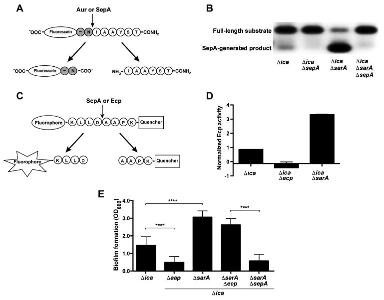
SarA negatively regulates SepA activity, Ecp activity, and SepA-dependent biofilm formation. A. Fluorescein-labelled peptide for activity of S. aureus Aur and S. epidermidis SepA. The cleavage leaves a negatively-charged, fluorescein-labelled species that can be observed by running the reaction on an agarose gel. B. Activity assay for SepA. Cell-free spent media from S. epidermidis strains was incubated with the fluorescein-labeled substrate. Reactions were run on an agarose gel and visualized under UV light. The negative image of the gel is shown. C. FRET (fluorescence resonance energy transfer)-based substrate for S. aureus ScpA and S. epidermidis Ecp. The peptide has an N-terminal fluorophore and C-terminal quencher linked to the terminal Lys residues. Cleavage allows fluorescence by separating the fluorophore from the quencher. D. Activity assay for Ecp. Cell-free spent media from S. epidermidis strains were incubated with the FRET substrate. Reaction rates (fluorescence/time) were averaged from two experiments with three replicates each and normalized to wild type 1457. E. Microtiter biofilm assay. Results are pooled from three experiments with three replicates each. One-way ANOVA with multiple comparisons (Bonferroni correction) was performed. **** indicates p < 0.0001.
