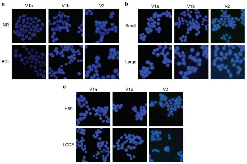
Figure 2.
By immunofluorescence, cholangiocytes from Normal and BDL rats (a) express V2 receptor subtype. After BDL, cells present low expression of V1a and V1b but higher immunopositivity for V2. The same experiments were performed in small and large cholangiocytes from mice (b), where the expression of V2 is predominant both in small and large cells. Finally, in human cholangiocytes (c), the immunofluorescence demonstrated that they express V2 with greater amount in LCDE, the cells from the cystic epithelium. Specific immunoreactivity of representative fields is shown in green; cell nuclei were stained with DAPI (blue). No staining was visible when primary antibodies were replaced with non-immune serum (data not shown). Bar = 200 μm.

