
Figure 4. Precocious suppression of transcytosis accelerates functional BRB formation.
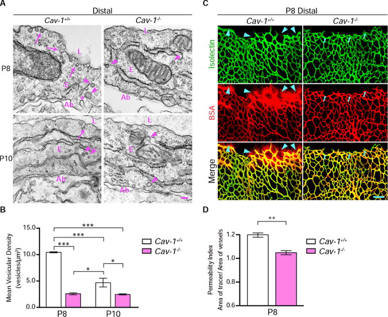
(A) Precocious suppression of transcytosis is observed in Cav-1−/&minus retinas. EM in distal vessels at P8 (top) reveals many vesicles associated with luminal (arrows) and abluminal membranes (*) and in the cytoplasm (arrowheads) in Cav-1+/+ endothelial cells. Cav-1−/&minus endothelial cells have drastically reduced numbers of vesicles. At P10 (bottom), few vesicles are observed in both Cav-1+/+ and Cav-1−/&minus distal vessels. (B) Quantifications of vesicles in distal vessels from Cav-1+/+ and Cav-1−/&minus mice at P8 and P10. Data are presented as mean ± s.e.m. (n= 3 mice per age per genotype, 15-20 vessels analyzed per mouse). Statistical significance was determined by two-way ANOVA with a post-hoc Bonferroni multiple comparison adjustment. (C) Intravenous injection of BSA (red) in P8 pups reveals tracer leakage in the retinal parenchyma from distal vessels of Cav-1+/+ (arrowheads) but not Cav-1−/&minus mice. (D) Permeability index from distal regions of P8 Cav-1+/+ and Cav-1−/&minus littermates. Data are presented as mean ± s.e.m. (n=3-4 mice per genotype). Statistical significance was determined by unpaired t-test. *, P < 0.05; **, P < 0.01; ***, P < 0.001. L – lumen, E – endothelial cells, Ab – abluminal. Scale bar represents 100 nm in (A) and 100 μm in (C).