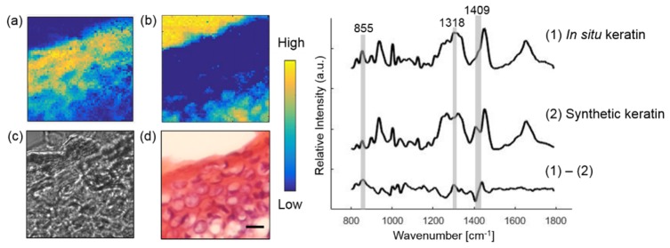
Fig. 3.
Extracting the epidermal component from a normal skin section. Raman images of in situ keratin (a) and Raman substrate (b) are compared with bright-field image (c) and histopathology image (d). Plots on the right show Raman spectra of in situ, synthetic keratin and their difference spectrum. Scale bar:10 μm.

