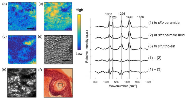
Fig. 5.
Extracting lipids within a hair follicle from a SCC skin section. In situ ceramide (a), triolein (b) and Raman substrate (c) are resolved from the image. Raman images are compared with the bright-field image (d), CLSM image (e) and histopathology image (f). Some lipids in (f) were lost during the staining processing. The box on (e) and (f) marks the location of Raman imaging. Difference spectrum between in situ ceramide and palmitic acid and difference spectrum between in situ ceramide and triolein are also shown. Scale bar: 10 μm.

