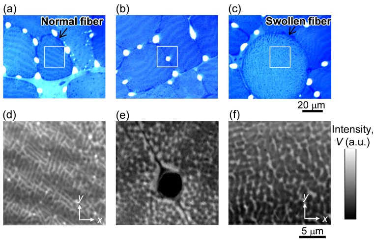
Fig. 3.
Images of stained tissues of muscle fibers for 1 day after excising. (a-c) Bright field images of three different areas. (d-f) PT images taken on the square area on (a-c), corresponding to the area at the center of the normal fiber (d), near the capillary (e), and at the center of the swollen fiber (f). Pump and probe laser powers are 50 μW and 1 mW, respectively.

