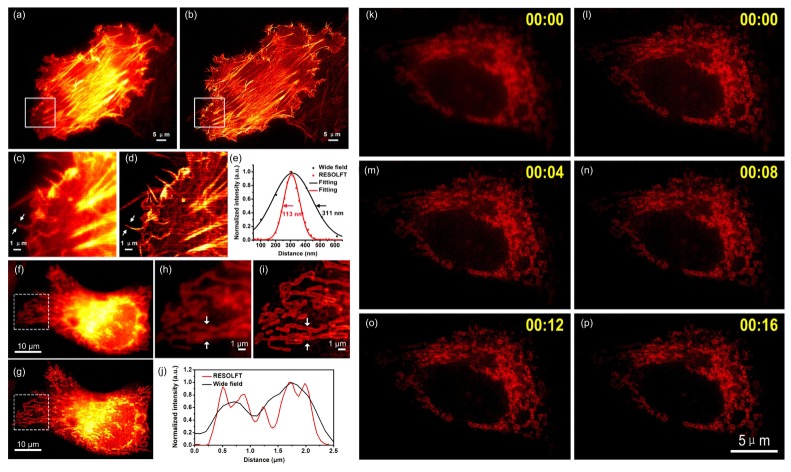
Fig. 4.
Super-resolution imaging of protein-protein interactions by combing BiFC with parallelized RESOLFT in live HeLa cells. HeLa cells co-transfected with Lifeact-rsEGFP2-N and Lifeact-rsEGFP2-C or Bak-rsEGFP2-N and Bcl-xL-rsEGFP2-C were imaged with a parallelized RESOLFT microscope. (a) Conventional wide-field fluorescence image and (b) RESOLFT image of cells expressing reconstituted rsEGFP2 signal mediated by Lifeact-rsEGFP2-N and Lifeact-rsEGFP2-C homodimerization. (c) The magnified image of boxed area of (a) and (d) the magnified image of boxed area of (b) are presented and intensity profiles measured of arrowed regions in (c) and (d) are presented in (e) with black and red curves, respectively. (f) Conventional wide-field image and (g) RESOLFT image of cells expressing reconstituted rsEGFP2 signal mediated by Bcl-xL and Bak heterodimerization. (h) The magnified image of boxed area of (f) and (i) the magnified image of boxed area of (g) are presented and intensity profiles measured of arrowed regions in (h) and (i) are presented in (j) with black and red curves, respectively. (k) Conventional wide-field image of a HeLa cell expressing reconstituted rsEGFP2 signal mediated by Bcl-xL and Bak heterodimerization. (l-p) Continuous time-lapse imaging of the HeLa cell in (k) with parallelized RESOLFT. A 405 nm continuous wave diode laser was used for on-switching (2 ms; 12 mW measured at the back focal plane of the objective, corresponding to 0.1 kW/cm2); a 488 nm continuous wave laser was used for off-switching (20 ms; 50 mW measured at the back focal plane of the objective, corresponding to 1 kW/cm2) and fluorescence readout (4 ms; 50 mW measured at the back focal plane of the objective). RESOLFT images were taken using a 24 nm scanning step (pixel) size and (360/24)2 steps were required for one RESOLFT frame in (b) and 36 nm scanning step (pixel) size and (360/36)2 steps were required for one RESOLFT frame in (g) and (l-p). Each RESOLFT frame was taken within ∼6s in (b) and ∼3s in (g) and (l-p). Scale bar: 5μm in (a) and (b),10μm in (f) and (g) and 1μm in (c),(d),(h),(i) and 5μm in (k-p). The images are displayed using linear intensity scale without adjusting γ factor. All the RESOLFT images are raw images without deconvolution.

