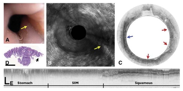
Fig. 12.
Presumed Barrett’s esophagus. (A) Videoendoscopic image shows an irregular SCJ with a tongue of healthy mucosa (arrow). (B) An MIP rendering of the entire volumetric OFDI data set, obtained at the corresponding location in A. (C) Corresponding cross-sectional OFDI image demonstrates squamous mucosa (blue arrow) interspersed with regions that satisfy the OCT criteria for BE (red arrows). (D) Histopathologic image of the biopsy specimen taken from the SCJ with no signs of intestinal metaplasia (H&E, orig. mag. 2). (E) A longitudinal OFDI cross-section through the SCJ shows a 12-mm segment of mucosa that is consistent with BE. Scale bars and tick marks represent 1 mm. (Figure and caption adapted from [129], with permission from Elsevier.)

