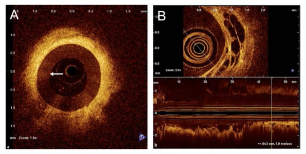
Fig. 19.
(A) Radial OCT image of the common bile duct in a patient with a benign stricture following cholecystectomy. The probe is surrounded by the endoscopic retrograde cholangiopancreatography catheter (arrow). (B) OCT criteria - large, non-reflective areas contained in the intermediate layer - suggesting tumor vessels. Both axial section and longitudinal reconstruction are depicted. (Figure and caption adapted from [154], by permission from Macmillan Publishers Ltd.)

