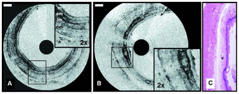
Fig. 2.
Images obtained using the first catheter-based time domain OCT system for visualizing the rabbit esophagus in vivo displayed in inverse gray scale. Esophageal layers can be clearly delineated: mucosa (m); submucosa (sm); inner and outer muscularis (im and om); serosa (s). A vessel (v) in the submucosa can also be seen. Reprinted from [29] with permission from Elsevier.

