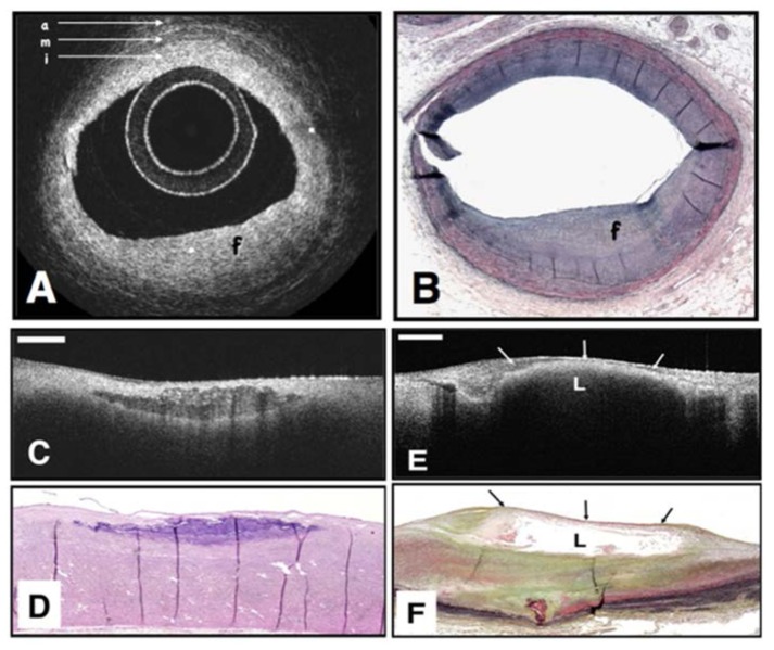
Fig. 3.
OCT image (A) of a fibrous coronary plaque showing a homogeneous, signal-rich interior (f) as well as the three-layered structure of intima (i), media (m), adventitia (a). Corresponding histology (B). OCT image of fibrocalcific aortic plaque (C) showing a sharply delineated region with a signal-poor interior. Corresponding histology (D). OCT image of a lipid-rich plaque (E) showing signal-poor region (L) with poorly delineated borders. Corresponding histology (F). Reprinted from [31].

