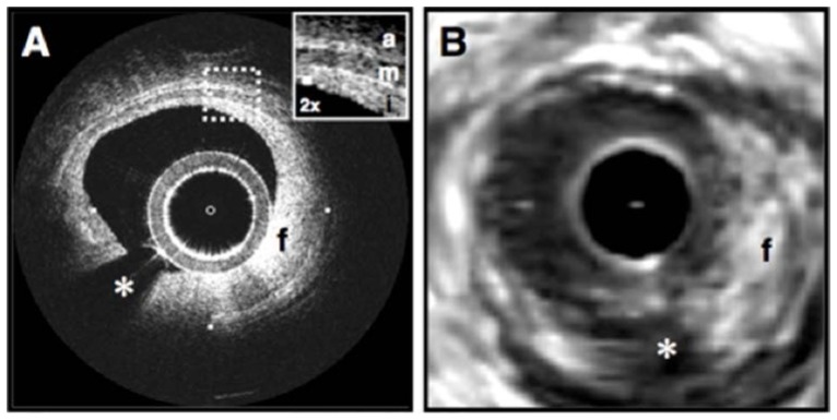
Fig. 6.
Fibrous coronary plaque imaged in vivo by OCT (A) and IVUS (B). A. From 9 o'clock to 2 o'clock, this OCT image demonstrates visualization of the intima (i, with intimal hyperplasia), media (m), and adventitia (a). The internal and external elastic laminae are visible as signal-rich lines bordering the media (inset). A plaque extending from 2 o'clock to 9 o'clock contains a homogeneous, signal-rich region consistent with a fibrous plaque (f) that is partially obscured by a guide wire shadow artifact (*). B. In the corresponding IVUS image, the fibrous plaque (f) is also visualized. Tick marks, 1 mm. Reprinted from [47].

