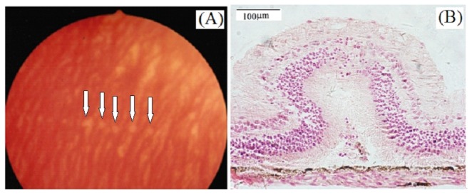
Fig. 5.
(A) Fundus photograph showing rabbit retinal lesions induced by pulsed 1338 nm laser. Photograph was taken at 24 hours post-exposure. The incident energy was 1.0 J. Arrows indicated the retinal lesions. (B) Histological section of retinal tissue at threshold level. Tissue was fixed at 48-h following laser exposures.

