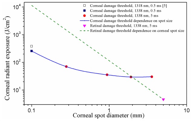
Fig. 6.
Corneal and retinal damage thresholds as a function of the incident corneal spot diameter for 5 ms pulsed 1338 nm laser. According to the action spectrum theory [3,8], the data point from Reference [5] could be adjusted to the data for 1338 nm, using the water absorption coefficients at the wavelengths of 1318 nm and 1338 nm (1.76 cm−1 for 1318 nm and 2.62 cm−1 for 1338 nm [23]). This point was plotted to illustrate the rapid change of damage threshold with the decrease of corneal spot size for small corneal spot sizes; for corneal spot diameters of 1 mm and above no significant spot size dependence could be observed. The green dashed line showed how the retinal damage threshold varied as the corneal spot diameter was decreased down from 5 mm, on the condition that the laser beam remained collimated and the damage threshold was expressed in corneal radiant exposure. The straight line was only an approximate analysis because the optical quality of the eye is much better in the central part than in outer parts and thus a smaller beam could well be focused better as compared to a larger beam, resulting in a smaller damage threshold.

