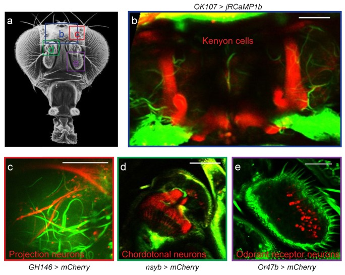
Fig. 6.
Transcutical imaging of Drosophila brain structures. (a) External view of a fly head. Image source: www.sdbonline.org. The four boxes denote imaging regions. The viewing angle may be slightly different for each region. (b) Mushroom body Kenyon cells of a fly bearing the OK107-GAL4 and UAS-jRCaMP1b transgenes. (c) Axon terminals of second-order projection neurons in the lateral horn region. Transgenes: GH146-GAL4 and UAS-mCherry. (e) Cell bodies of the Or47b odorant receptor neurons. Transgenes: Or47b-GAL4 and UAS-mCherry. (b-e) Red color: fluorescence signals from the labeled neurons. Green color: third harmonic signal. Scale bar, 50 µm.

