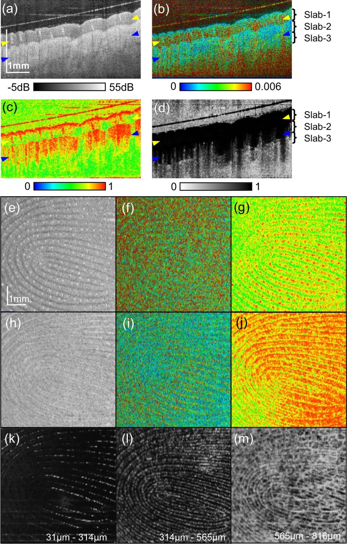
Fig. 5.
Multi-contrast images of finger pad. Cross-sectional (a) intensity, (b) birefringence tomography, (c) DOPU tomography, and (d) OCT-A. (e)–(g) are en face slices at the depth indicated by yellow arrowheads in the cross-sectional images. (h)–(j) are en face slices at the depth of blue arrowheads. (e) and (h): scattering intensity, (f) ang (i): birefringence, (g) and (j): DOPU. (k)–(m) are slab projections of OCT-A. The depth positions of the slabs are indicated in (b) and (d).

