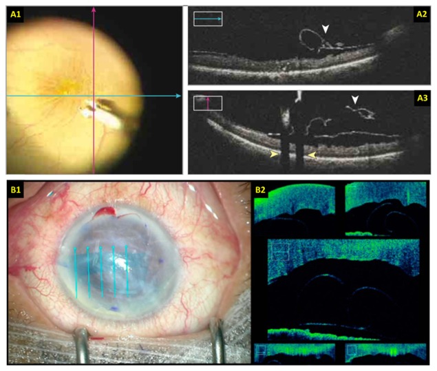
Fig. 6.
Surgical imaging with live 2D MIOCT. (A) Retinal surgery imaging protocol using two orthogonal high-resolution B-scans [118]. (B) Anterior eye surgery imaging protocol using five parallel and laterally offset B-scans spanning the area of interest [132]. The B-scan locations are overlaid on the surgical views.

