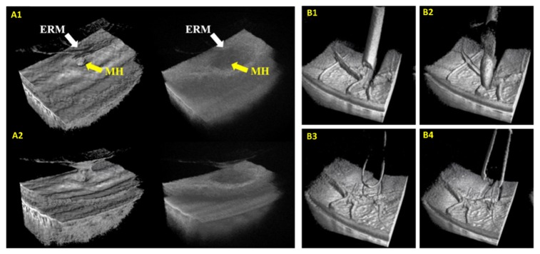
Fig. 7.
Surgical imaging with live 3D MIOCT. (A) Real-time enhanced volumetric rendering using GPU-accelerated software. Median filtering and depth-based shading improved visualization of subtle anatomical and pathological structures [137]. Manipulation of the rendering perspective also allowed surgeons to visualize structures from various perspectives. (B) Visualization of commonplace surgical instruments in OCT volumes acquired during porcine eye surgery. ERM: epiretinal membrane, MH: macular hole.

