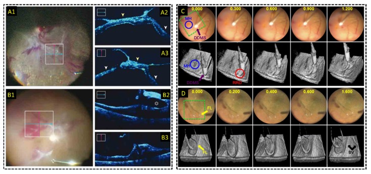
Fig. 12.
Live MIOCT imaging during human retinal surgery. (A-B) Live 2D MIOCT imaging with high-resolution orthogonal B-scans using the commercial Zeiss RESCAN 700 [118] during (A) retinal detachment and (B) proliferative diabetic retinopathy procedures. (C-D) Live 3D MIOCT imaging using real-time volumes [122] during retinal brushing with a (C) diamond dusted scraper and a (D) flex loop. High-speed volumetric imaging enhances visualization of subtle 3D tissue deformations.

