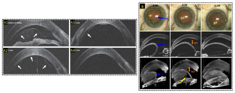
Fig. 13.
Live MIOCT imaging of human anterior eye surgery. (A) Live 2D MIOCT imaging with high-resolution B-scans using the commercial Haag-Streit Surgical iOCT [188] during Decemet’s membrane endothelial keratoplasty. Real-time direct visualization graft attachment during air filling in B-scans was made possible by MIOCT (B) Live 3D MIOCT imaging using real-time volumes [122] during Decemet’s stripping automated endothelial keratoplasty. Graft unfolding, orientation, and apposition was directly visible in the volumes.

