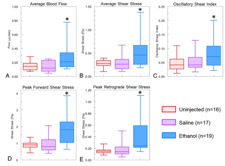
Fig. 3.
Hemodynamic measurements. Ethanol-exposed embryos exhibited higher levels of all hemodynamic measurements. (A) Average blood flow over a heartbeat from multiple B-scan images. (B) These same images were used to determine the average shear stress. Ethanol-exposed embryos had significantly higher average blood flow and shear stress. (C) The shear stress values over the heartbeat were used to determine the oscillatory shear index which was higher in ethanol-exposed embryos due to the higher retrograde blood flow. (D and E) the peak forward and retrograde shear stress were determined and shown to be higher in ethanol-exposed embryos in comparison with uninjected and saline controls. *indicates p < 0.05.

