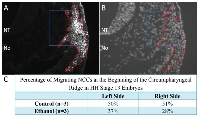
Fig. 5.
Quantification of cardiac neural crest mid-migration. Transverse cryosections of stage 13 embryos at the level of the cardiac neural crest were immunofluorescently stained using HNK-1 and anti-AP-2 antibodies to identify neural crest cells (A). Cyrosections were also co-stained with DAPI to identify all cell nuclei (B) for quantification. The area of HNK-1/AP-2 positive neural crest cells are outlined in red (A,B) with the area that was selected for quantification outlined in blue regions of interest (A,B). The area selected is within the beginning of the circumpharyngeal ridge. Both the left and right sides of the embryo were similarly imaged for quantification. NT = neural tube; No = notochord.

