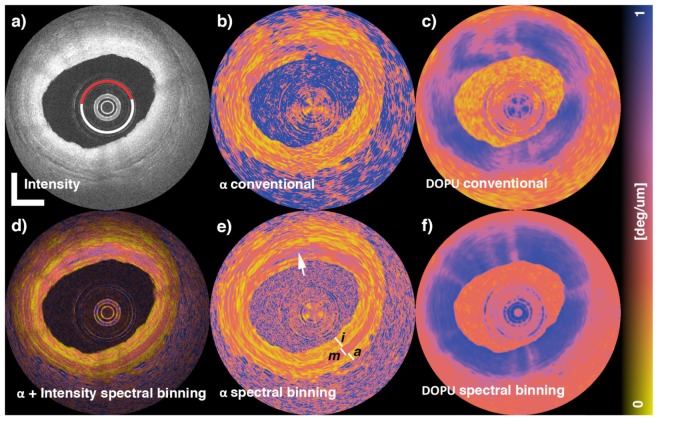
Fig. 10.
Cadaveric human coronary artery PS-OCT images with and without numerical non-common-path-PMD correction by a spectral binning method. The first row shows intensity, local retardation, DOPU without PMD correction (left to right). The second row shows intensity-local retardation composite, local retardation, DOPU with PMD correction (left to right.) The images were reprinted from [70].

