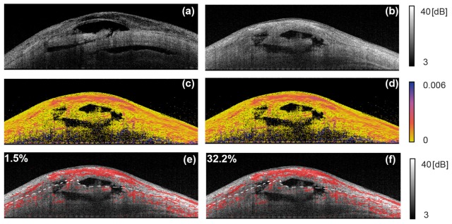
Fig. 12.
Examples of birefringence imaging of blebs. (a) and (b) are intensity OCT and (c) and (d) are birefringence cross-sections. In (e) and (f), the high birefringence pixels (red) are overlaid on intensity image. The numbers at the top left indicate the areal fraction of high birefringence regions in the conjunctiva (adapted from [85]).

