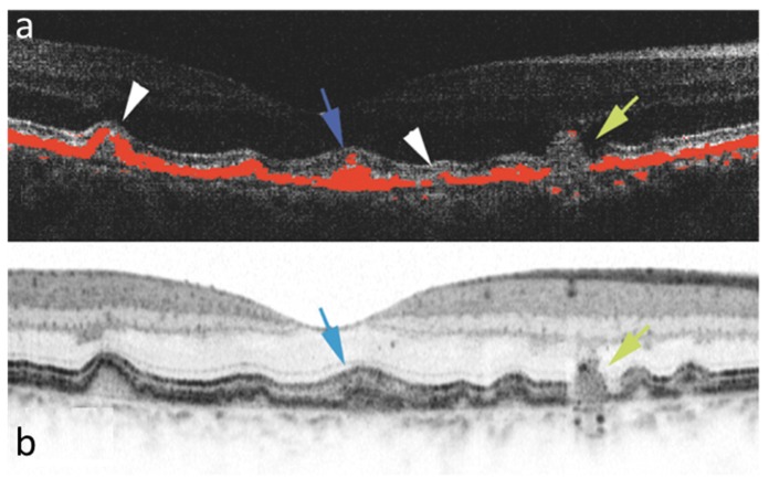
Fig. 17.
Drusen imaging by PS-OCT (a) and intensity based OCT (b) in the same eye of a patient with AMD. Various forms of drusen can be differentiated in the PS-OCT image, where depolarizing tissue (red) has been segmented by its low DOPU value. Blue arrow: druse filled with depolarizing material; white arrowheads: small atrophic lesions; yellow arrow: drusenoid structure with complete loss of RPE. (Reproduced from [119] by permission of the Association for Research in Vision and Ophthalmology)

