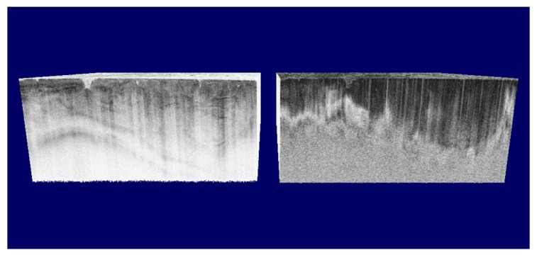
Fig. 20.
Structural intensity (left) and polarization sensitive image (right) of a hamster cheek pouch cancer model. In the intensity image the features of the tumor tissue are visible, however, the PS image shows significant contrast between normal (left) and tumor tissue (right) based on the banding pattern. The normal cheek pouch tissue shows a much higher birefringence than the cancer tissue. (Adapted from [57])

