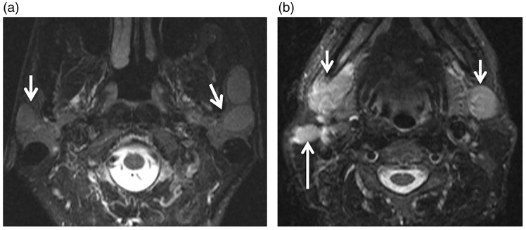
Figure 11.
Sarcoidosis. (a) Axial T2-weighted image shows well-defined hyperintense focal lesions (arrows) in both parotid glands more on the left side. (b) Axial T2-weighted image at another level shows enlarged both submandibular glands (short arrows) more on the right side with enlarged adjacent cervical lymph (long arrow).

