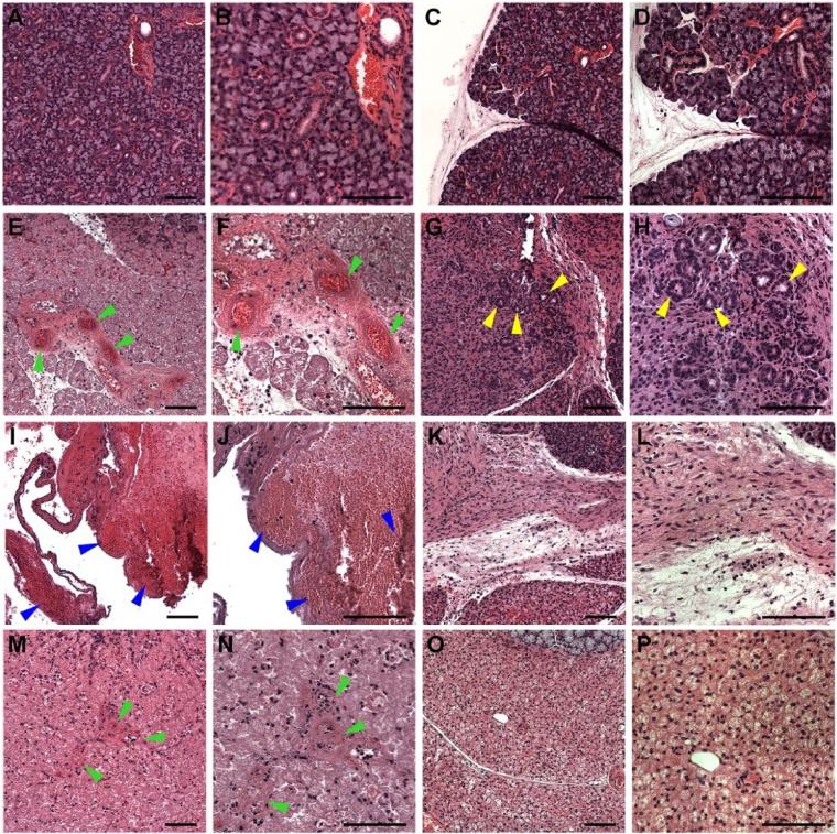
Figure 3.
Hematoxylin and eosin–stained images of mouse submandibular glands (mSMGs) treated with and without fibrin hydrogel (FH) scaffolds (magnification 10× or 20× from Appendix Fig. 3; yellow boxes). Shown are native mSMGs (A, B: day 0), wounded mSMGs without scaffold (C, D: day 0), wounded mSMGs with dye-conjugated FH scaffold with laminin 1 peptide (DyLight 680; E, F: day 3; G, H: day 8), wounded mSMGs without scaffold (I, J: day 3; K, L: day 8), and wounded mSMGs with dye-conjugated FH scaffold (DyLight 680; M, N: day 3; O, P: day 8). Arrowheads indicate blood vessels (green), organized round structures with lumen (yellow), and blood clots (blue), respectively. Scale bars represent 100 µm. Representative image from a total of 5 mice per group.

