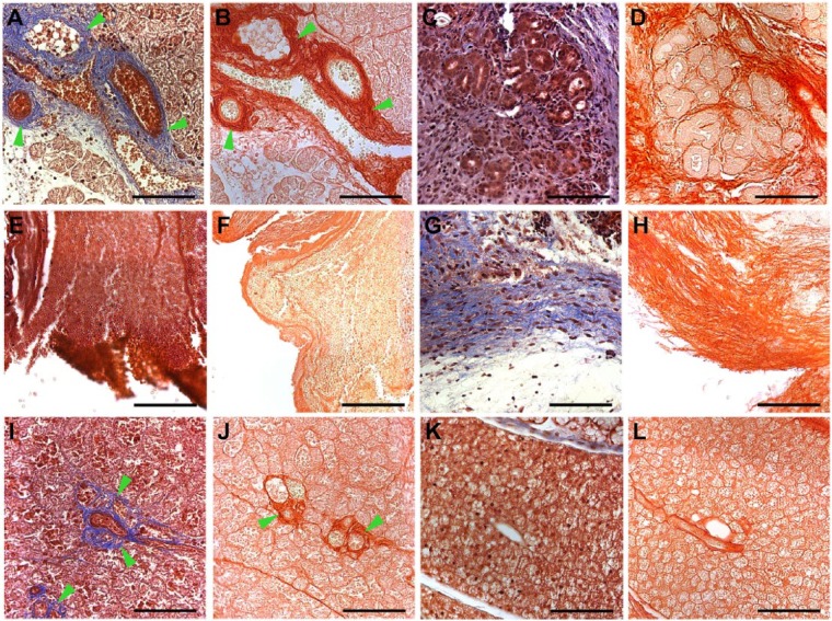
Figure 4.
Stained images of mouse submandibular glands (mSMGs) treated with and without fibrin hydrogel (FH) scaffolds (magnification 20×): Masson’s trichrome (A, C, E, G, I, K) and picrosirius red (B, D, F, H, J, L). Shown are wounded mSMGs with dye-conjugated FH scaffold with laminin 1 peptide (DyLight 680; A, B: day 3; C, D: day 8), wounded mSMGs without scaffold (E, F: day 3; G, H: day 8), and wounded mSMGs with dye-conjugated FH scaffold (DyLight 680; I, J: day 3: K, L: day 8). Scale bars represent 100 µm. Green arrowheads indicate collagen fibrils. Representative image from a total of 5 mice per group.

