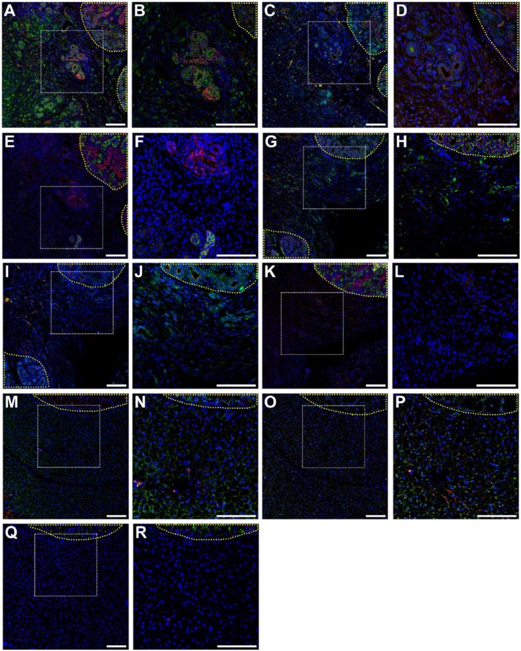
Figure 5.
Salivary structural and functional marker organization in wounded mouse submandibular glands (mSMGs) with dye-conjugated fibrin hydrogel (FH) scaffold with laminin 1 peptide (DyLight 680; A–F), wounded mSMGs without scaffold (G–L), and wounded mSMGs with dye-conjugated FH scaffold (DyLight 680; M–R) was determined with confocal microscopy (magnification 10x or 20x from white dotted areas) as follows: zonula occludens 1 (green; A, B, G, H, M, N), E-cadherin (red; A, B, G, H, M, N), TMEM16A (green; C, D, I, J, O, P), cytokeratin 7 (red; C, D, I, J, O, P), aquaporin 5 (green; E, F, K, L, Q, R), Na+/K+-ATPase (red; E, F, K, L, Q, R), and TO-PRO-3 (blue; everywhere). Yellow-dotted areas are unwounded areas. Scale bars represent 100 µm. Representative image from a total of 5 mice per group.

