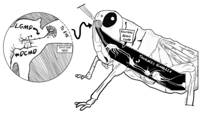
Figure 1.
Neuroanatomy of the grasshopper’s motion detector neurons. The grasshopper optic lobes (lamina, medulla and lobula) lie in the central brain beneath each of the compound eyes and processes visual information. The lobula giant motion detectors (LGMDs) receive visual signals from the lobula and pass these inputs to the descending contralateral motion detectors (DCMDs). The LGMDs respond to motion of an object seen moving across the insect eye as well as to the looming effect, where an object increases in size as it approaches the eye. The DCMD activates motor neurons in the thoracic ganglia. These primary motion detector neurons underpin the animal’s detector and motor response to a looming object, forming the see-and-jump escape mechanism.

