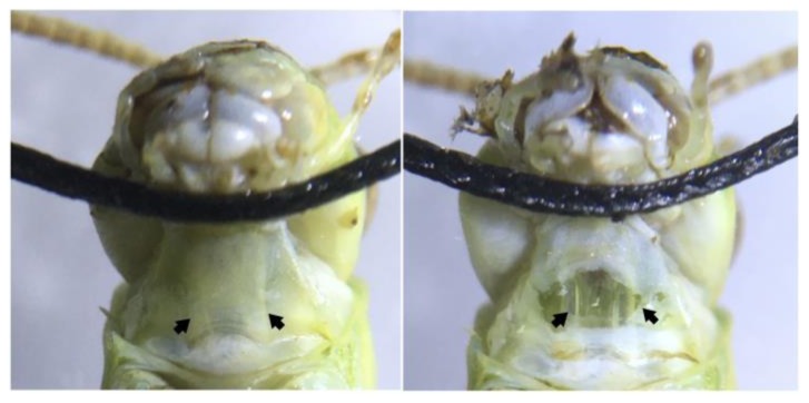
Figure 2.
Ventral view of the grasshopper neck showing the neck connectives from the head to the thoracic ganglia. (Left) Neck connectives (pointed to by arrows) are visible as white stripes under the neck skin. Sewing thread is used to hold the grasshopper’s head back and in place during the experiments. (Right) A simple neck cut exposes the connectives, two translucent tubes, where the part of the ventral nerve cord can be found. Placing a hook electrode around one connective and exposing the contralateral eye to looming visual stimuli allows DCMD activity to be recorded.

