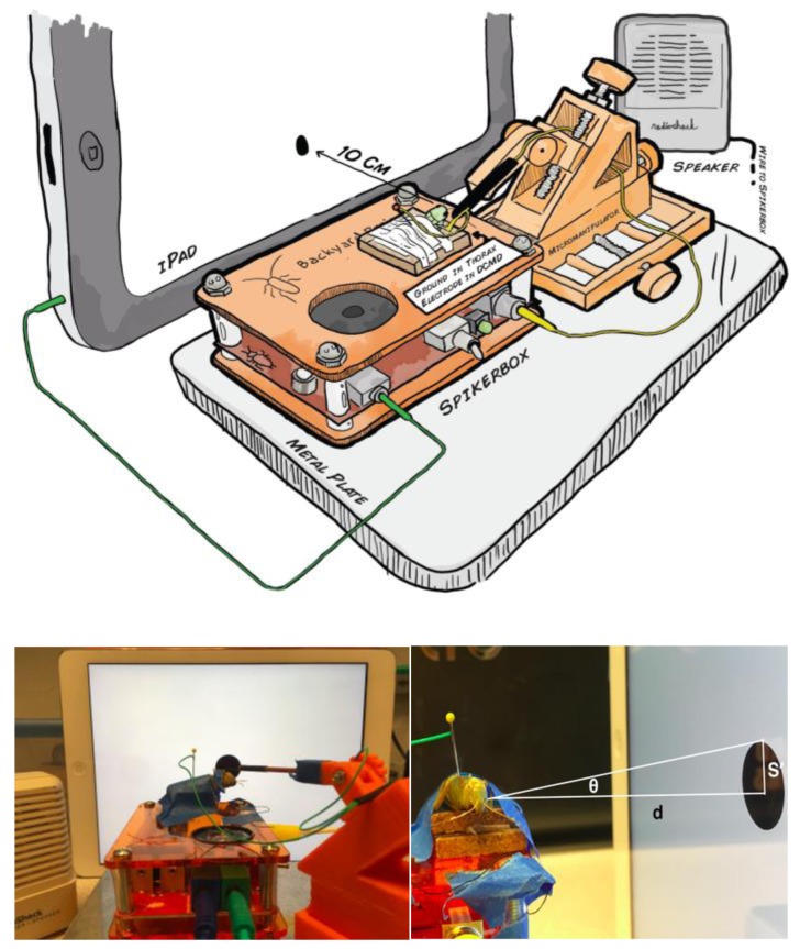
Figure 3.
Experimental setup to measure descending contralateral movement detector (DCMD) activity during simulated stimulus presentation. (Top) Schematic representation of the experimental setup, grasshopper is taped down onto the corkboard on top of the SpikerBox. The reference electrode is grounded at the thorax and the recording hook electrode, guided by the 3D printed micromanipulator for precision, is placed around the neck connective where the ventral nerve cord and DCMD neuron’s axon is found. The iPad presenting the visual stimuli is placed 10cm from the grasshopper’s eye and the angle between the grasshopper’s eye and the center of the iPad is minimized. (Bottom left) Photograph of the apparatus. (Bottom right) Visual stimuli are displayed on the iPad to simulate the looming effect of an object approaching the grasshopper. The radius (S′) and subtending angle (θ) of a filled black dot can be manipulated to simulate a fixed sized object (S) approaching at a particular constant velocity (v) towards the eye of the insect a fixed distance (d) from the screen. DCMD recordings can be made during visual simulations of different sized objects moving at different velocities.

