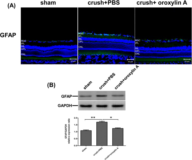
Fig 6. Oroxylin A attenuated retinal gliosis at 2 weeks after ON crush.
(A) GFAP (astrocytes and Muller cells) immunoreactivity in retinal sections. Effects of oroxylin A on the suppression of GFAP level in the retina at 2 weeks after ON crush. (B) Western blotting showing the expression levels of GFAP in the retina. In the bar graph, the expression level of GFAP is expressed as a ratio to GAPDH expression Values for sham-operated retinas were set to 1. Results represent the means ± S.D for three independent experiments. *p<0.05. **p<0.01. Scale bar: 50μm.

