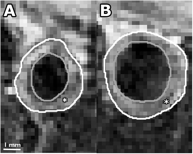
Fig 3.
Contrast-enhanced T1w BB MR image of atherosclerotic plaque for a non-treated rabbit (panel A) and Ivabradine treated rabbit (panel B). In the panels, the outer vessel wall is indicated by the white line and the lumen by the gray line. The atherosclerotic plaque is indicated by the asterisk (*).

