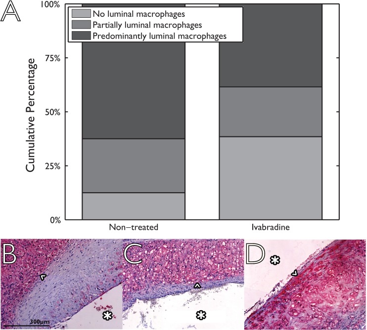
Fig 5. Presence of macrophages in the proximity of the vascular lumen using a semi-quantitative three point scale for non-treated (eight animals, two sections per animal) and Ivabradine-treated animals (seven animals, 2 sections per animal).
Histological sections are divided into three categories ranging from 0 (no luminal macrophages; example image shown in panel B) via 1 (partially luminal macrophages; example in panel C) to 2 (predominantly luminal macrophages; example in panel D). Macrophages in the histological images are indicated by an arrow head (^). The displayed scale bar is applicable for all histological images and the luminal side is indicated by an asterisk (*) in all histological images.

