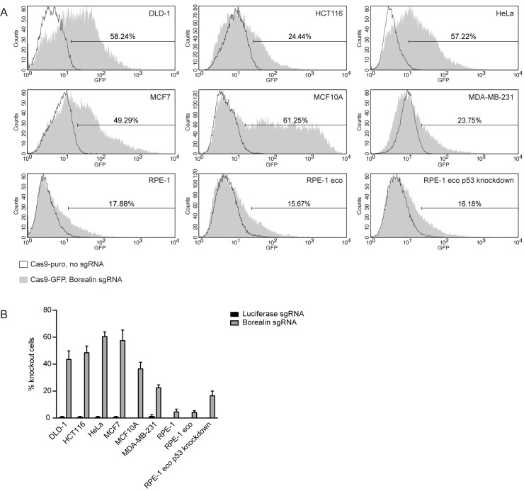
Fig 3. Effect of CRISPR/Cas9 baculoviral transduction in a panel of cell lines.
A) Representative FACS-profiles showing GFP expression in cells treated with Cas9-GFP baculovirus (MOI: 75). The markers are set such that 2% of the cells treated with Cas9-puro baculovirus are included in this region. The percentage of cells treated with Cas9-GFP baculovirus within the marker region is indicated. B) Immunofluorescence images of mitotic cells treated with the indicated Cas9-GFP baculoviruses (MOI: 75) were scored by eye for the presence or absence of centromeric Aurora B. The bars represent the mean ± SD of 2 experiments. At least 24 cells were analyzed per experiment per condition.

