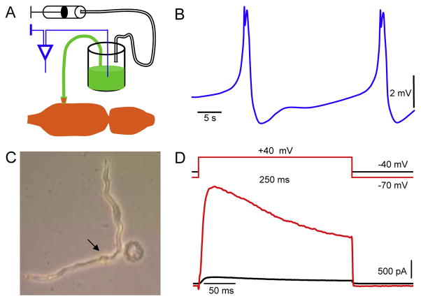
Fig. 3.
Whole heart and single cardiomyocyte recordings. A. The schematic drawings of experimental approach for the recording of cardiac activity using the suction electrode. Note that the electrode container is filled with 150 mM KCl solution supplemented with dye in order to facilitate the visual ablation and to prevent the myocardial damage. B. The ventricular APs that underlie each heartbeat. C. Snapshot of enzymatically isolated ventricular cardiomyocyte. The arrow points to the nucleus of the healthy cell, and the damaged cell is round. D. Family of outward K+ currents in cardiomyocyte activated from different holding potentials. Scale bars are shown.

