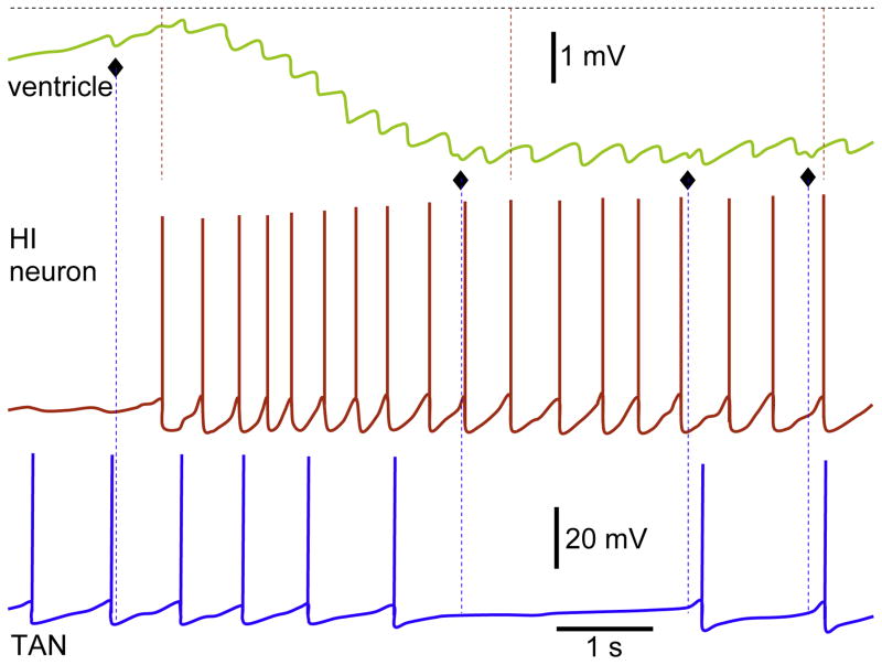
Fig. 4.
Simultaneous recordings from ventricle and two neurons. The IPSPs in ventricle are derived by APs in HI neuron. During recording, four additional IPSPs (屦) were observed, which were not mediated by HI and TAN. Note similar latencies between the APs in HI and corresponding IPSPs in the ventricle; these are shown with dashed red lines for randomly chosen responses. The amplitude scale bar for both neurons and time scale bar for all traces are identical.
Reproduced with permission and modified from Fig. 2 (Bychkov et al., 1997).

