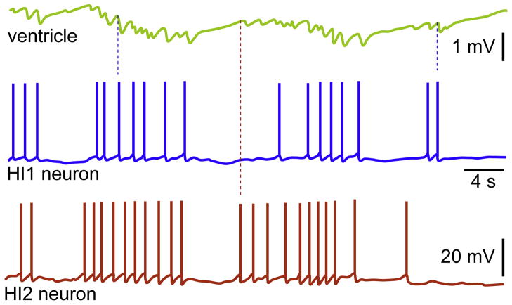
Fig. 5.
Spontaneous activity in heart inhibitory neurons. The properties of membrane potential and APs were simultaneously analyzed in ventricle and neurons, HI1 and HI2. Note that the burst activity to some extent coincides in HI1 and HI2, which subsequently results in summation of IPSPs. The dashed blue and red lines indicate randomly chosen APs in HI1 and HI2, respectively; they also refer to corresponding IPSPs and latencies after each APs.
Reproduced with permission (Bychkov et al., 1997).

