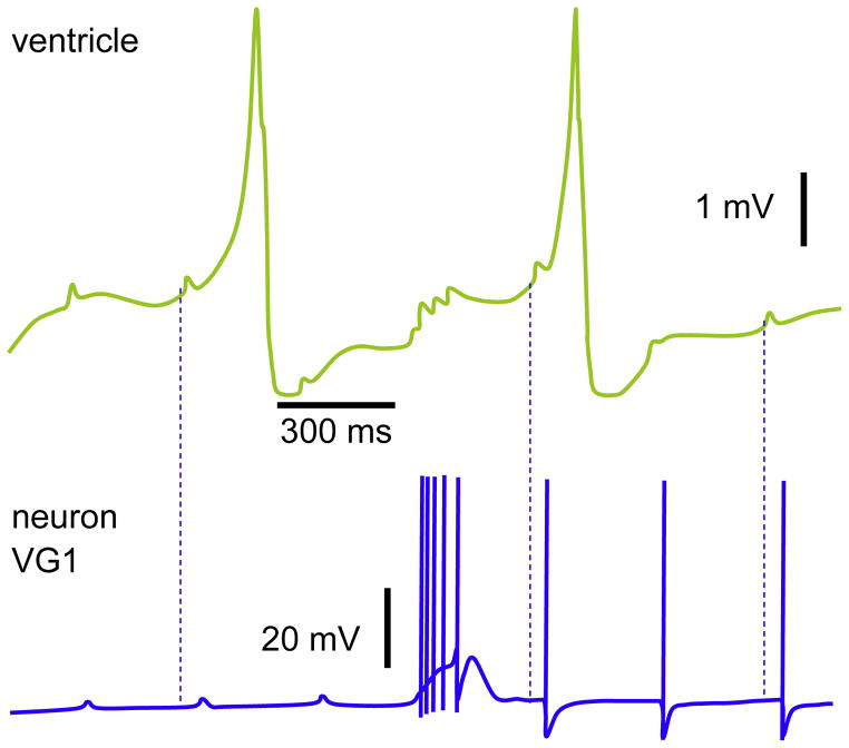
Fig. 8.
Cardioregulatory neuron VG1. The EPSPs in the ventricle and antidromic APs (the three last) were evoked by the direct stimulations of heart. The spontaneous burst coincides with the current injections applied to the ventricle. Note the resultant EPSPs in the ventricle after each AP within the burst. Dashed lines indicate the latency of VG1 response to EPSPs in ventricle; the corresponding pairs are randomly chosen.
Reproduced with permission (Zhuravlev et al., 1997).

