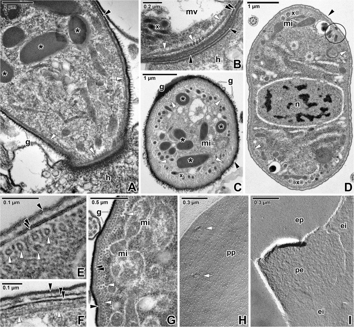
Fig 2. Cortex organisation in Siedleckia nematoides.
A. Apical end of a parasite attached to the host enterocyte. Note the well-developed layer of glycocalyx. B. A detail of parasite apical end focusing on organisation of subpellicular microtubules. C. General view of parasite cross-sectioned in the anterior region. D. General view of a parasite cross-sectioned in the middle region. E. The cross-sectioned pellicle with well-preserved and adjacent cortical cytomembranes. F. Longitudinally-sectioned pellicle with obviously separated cortical cytomembranes. G. Cortex of parasite cross-sectioned in the middle region. H. Protoplasmic fracture face of the plasma membrane with pores. I. Fractured plasma membrane and cortical cytomembranes. A, C, G: RR TEM; B, D-F: TEM; H-I: FE TEM. black arrowhead–plasma membrane, black asterisk–rhoptry, black circle–pore, double/paired black arrowhead–IMC, ei–EF of the internal cytomembrane, ep–EF of the plasma membrane, g–glycocalyx, h–host tissue, mi–mitochondria, mv–mucronal vacuole, n–nucleus, pe–PF of the external cytomembrane, pp–PF of the plasma membrane, white arrowhead–subpellicular microtubule, white arrows–pores, x—micronemes.

