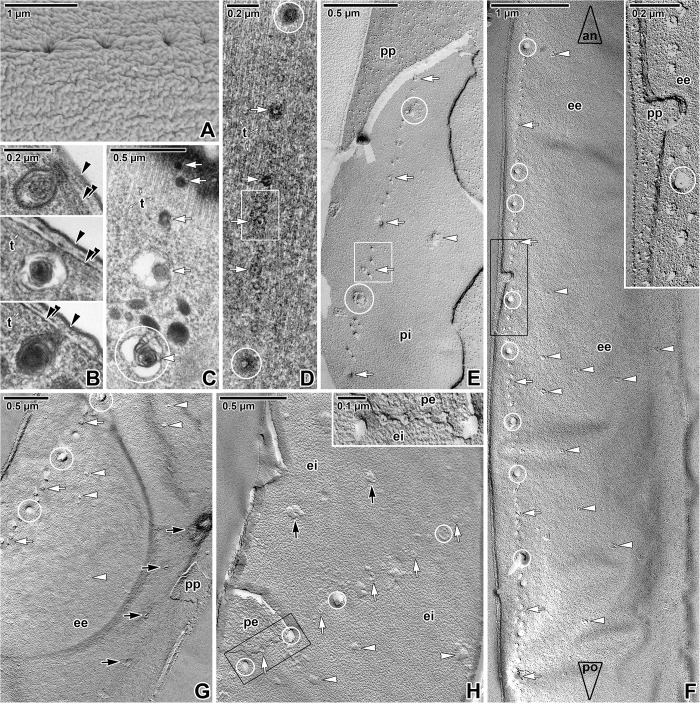
Fig 3. Distribution of the pores on the Siedleckia nematoides surface.
A. Detail of pellicle surface with a well visible row of pores. B. Different longitudinally-sectioned vesicular structures connected to the pellicle and corresponding to the pores observed by SEM. C. An almost superficial section of a parasite revealing the pores and vesicles organised in row. D. Superficially-sectioned cortex showing the layer of subpellicular microtubules and a row of pores of various size. E. Fractured pellicle revealing the row of differently sized pores located on the PF of the internal cytomembrane, but not visible at the plasma membrane. F. A general view of the longitudinally fractured pellicle revealing the external cytomembrane with a lateral row of pores and few randomly distributed pores. The large empty arrowheads with labels show the direction towards anterior (an) and posterior (po) parasite ends. The inset shows the fractured pellicle and pores demarcated by black rectangle in more detail. G. Fractured pellicle showing pores organised in rows; few pores are distributed randomly. H. A fragment of fractured pellicle where several rows of variously sized pores are visible. Inset shows a more detailed view of area demarcated by black rectangle, with alternating small and large pores organised in row. A: SEM; B, D: TEM; C: RR TEM; E-H: FE TEM. black arrowhead–plasma membrane, black arrows–additional row of pores, double/paired black arrowhead–IMC, ee–EF of the external cytomembrane, ei–EF of the internal cytomembrane, pe–PF of the external cytomembrane, pi–PF of the internal cytomembrane, pp–PF of the plasma membrane, t–subpellicular microtubules, white arrows–lateral row of pores, white arrowheads–randomly distributed pores, white circles indicate some of the large pores, white rectangle demarcates the doubled row of pores.

