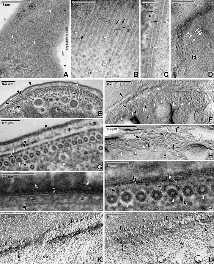
Fig 4. Organisation of the subpellicular microtubules in Siedleckia nematoides.
A. A superficial section of a cortex revealing the pores and subpellicular microtubules being helically twisted along the longitudinal cell axis. B-C. Higher magnification of the longitudinally-sectioned subpellicular microtubules. Note the rows of filamentous structures running parallel to the adjacent microtubules (grey arrows) and filamentous connections with the microtubules (black arrows). D. Cytoplasmic face of the internal cytomembrane with IMP alignments (white arrows) that correspond to the localisation of subpellicular microtubules. E. The pellicle covering the anterior part of parasite, underlain by one continuous and several intermittent layers of subpellicular microtubules sectioned in cross (left) and tangential (right) plane. F. The view (similar to E) of fractured pellicle underlain with several layers of subpellicular microtubules. G. The cross-sectioned cortex in the middle region of parasite, showing the organisation of subpellicular microtubules with cross-linking protein complexes. H. Fractured subpellicular microtubules with cross-linking protein complexes. I. The detail of pellicle covered by a thick glycocalyx layer. J. The high magnification of cross-sectioned microtubules partially revealing the organisation of tubulin protofilaments. K-L. Various views of fractured pellicle revealing the cross-linking protein complexes. A, E, G, J: TEM; B-C, I: RR TEM; D, F, H, K-L: FE TEM. black arrow–filamentous structures around subpellicular microtubules, black arrowhead–plasma membrane, c–cytoplasm, double/paired black arrowhead–IMC, g–glycocalyx, grey arrow–filamentous structures located between individual microtubules, grey arrowhead–protein complexes localised between the plasma membrane and IMC, iti–inner surface of the true (= not fractured) internal cytomembrane, white arrow–protein complex embedded in the IMC, white arrowhead–subpellicular microtubule. Black circles mark some of the large pores. White ellipse encircles the cross-linking protein complexes anchoring the subpellicular microtubules to the internal cytomembrane.

