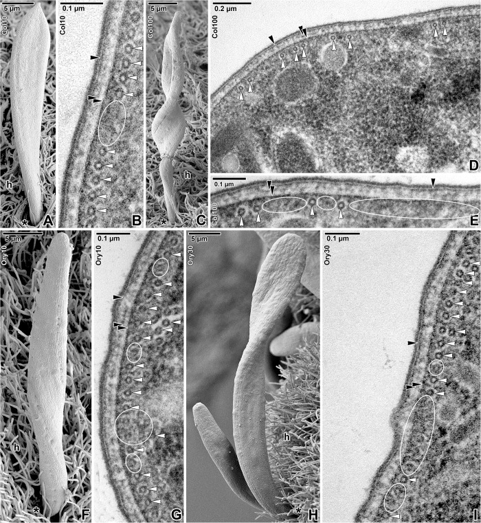
Fig 5. Organisation of the subpellicular microtubules in Siedleckia nematoides after treatment with cytoskeletal drugs.
A-B. Treatment with 10 mM colchicine for 2 h: A. Attached gamont. B. Cross-sectioned cortex with subpellicular microtubules. C-E. Treatment with 100 mM colchicine for 1 h: C. Attached gamont. D-E. General view (D) and higher magnification (E) of the cross-sectioned cortex with subpellicular microtubules. F-G. Treatment with 10 μM oryzalin for 8 h: F. Attached gamont. G. Cross-sectioned cortex with subpellicular microtubules. H-I. Treatment with 30 μM oryzalin for 7 h: H. Attached trophozoite and gamont. I. Cross-sectioned cortex with subpellicular microtubules. A, C, F, H: SEM; B, D-E, G, I: TEM. black asterisk–parasite apical end, black arrowhead–plasma membrane, double/paired black arrowhead–IMC, h–host tissue, white arrowhead–subpellicular microtubule. White ellipses demarcate the regions with disrupted microtubules.

