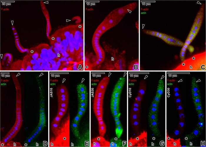
Fig 8. Phalloidin (TRITC) and antibody (FITC) staining of actin in Siedleckia nematoides before and after application of cytoskeletal drugs.
A-D. Non-treated parasites: A-B. Localisation of F-actin with phalloidin in parasites attached to the host tissue. C. Double labelling with phalloidin and specific anti-actin antibody. D. Double labelling with phalloidin (left) and anti-actin antibody (right), image split into two separate channels. E-F. Double labelling with phalloidin (left) and anti-actin antibody (right) in parasites treated with JAS, images split into two separate channels: E. 10 μM JAS (8 h). F. 30 μM JAS (6 h). G-H. Double labelling with phalloidin (left) and anti-actin antibody (right) in parasites treated with cytochalasin D, images split into two separate channels: G. 10 μM cytochalasin D (9 h). H. 30 μM cytochalasin D (8 h). A-B, D-H left: CLSM, phalloidin-TRITC/Hoechst; C: CLSM, IFA/phalloidin-TRITC/Hoechst; D-H right: CLSM, IFA/Hoechst; A-H: PFA fixation. black arrowhead–parasite caudal end, black asterisk–parasite apical end, h–host tissue.

