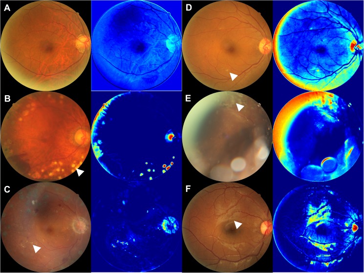
Fig 4. The images from the middle layer of the neural network.
(A) Representative color fundus photograph of NDR and an image of the middle layer, which has few characteristics. B-F: Representative color fundus photographs of PDR and their images of the middle layer. (B) Laser scars (white arrow head) were enhanced in the middle image. (C) Hard exudates (white arrow head) were enhanced in the middle image. (D) Soft exudates (white arrow head) were enhanced in the middle image. (E) Proliferative membranes (white arrow head) were enhanced in the middle image. (F) Reflections of the retina (white arrow head) were enhanced in the middle image.

